screening innovations

Screening Innovations: Opportunity in Perspective

If the history of medical innovation has taught us anything, the technology that will replace the traditional screening mammogram has most likely already been invented. You just can’t expect that anyone will recommend it to you, nor should you expect insurance to cover it. Years of clinical study and millions of dollars will be needed to verify the results, convince referring doctors and assure insurance coverage.

And, if you think that hospitals, imaging centers and all breast experts will enthusiastically and rapidly embrace the new technology when mammography’s replacement is clinically proven, you’d be sadly mistaken. There is up to a 17 year lag in the adoption of new medical devices, even when they are clinically proven.(1)

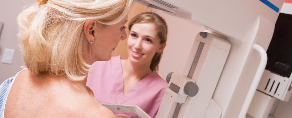
If you were to ask most doctors their opinion of the best breast cancer screening tool, most would say—without hesitation—the mammogram. They say this because they have been told that mammography is the only screening technology that has been proven to save lives in numerous clinical studies. But that doesn’t mean other screening technologies aren’t going to be shown to be better at saving lives… or shown to provide superior detection when used in conjunction with mammography. Other technologies just aren’t yet supported by as many clinical studies as the mammogram, which is now a 40 year-old technology. Clinical studies are extremely expensive and take much time, but we are able to evaluate their promise based on the few studies that are available or in progress, practitioner experience with the technology and a little bit of common sense.

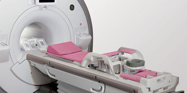
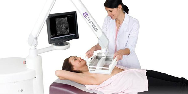
Here are the technologies that are either challenging the traditional mammogram as the gold standard, or that are used in conjunction with mammography to provide a greater chance for early detection and/or improve diagnostic capabilities:

Anatomical Imaging Tests

–––––––––––––––––––––––––––––––––––––––––––––––––––

Anatomical tests produce images of the body’s internal anatomy. The advantages of these tests is that they are typically covered by insurance, less expensive to conduct and are widely available. The disadvantages are that they cannot detect the metabolic activity within the body and/or are highly dependent on the skills of the individual administering or interpreting the test.

TOMOSYNTHESIS

–––––––––––––––––––––––––––––––––––––––––––––––––––

Breast tomosynthesis, known to most as 3D mammography, has been available commercially outside the United States since 2008. The FDA approved tomosynthesis in 2011. Numerous European and American clinical studies have demonstrated that adding tomosynthesis to a screening mammogram increases the cancer detection rate by about 40% and significantly lowers recall rates.(2) Tomosynthesis is a profound innovation that is improving the mammogram as a breast cancer screening tool.

 But will you be having one? Years after FDA approval, the majority of imaging centers and hospitals who offer screening mammograms do not offer tomosynthesis. Adoption also varies by region, with the percentage of practices using it in the northeastern region at 36.9% while the adoption rate in the south is just 18.4%.(3)

fast breast mri
FAST BREAST MRI

–––––––––––––––––––––––––––––––––––––––––––––––––––

On August 1, 2014, the Journal of Clinical Oncology published a proof-of-concept study. Fast Breast MRI, a three-minute MRI screening protocol for breast cancer, was shown to be as good as a regular breast MRI (that takes 21 minutes) and more accurate than digital mammography. It detected 11 invasive breast cancers that escaped detection on regular mammography, according to Christiane A. Kuhl, MD from the University Hospital of Aachen in Bonn, Germany. The researchers also found that although the test was highly sensitive, over-diagnosis was not an issue. FAST breast MRI “is a huge step forward in breast cancer screening,” according to Elizabeth A. Morris, MD, chief of breast imaging services at the Memorial Sloan Kettering Cancer Center in New York City. “Data clearly demonstrate that FAST breast MRI could be the standard for breast cancer screening. It is safe, does not induce cancers, and can find more cancers than mammography.”

The proof-of-concept study consisted of 443 women who were at mild to moderate risk for breast cancer. Each had a digital mammogram, and the women within the group who had dense breasts had also undergone ultrasound screening.  Yet, the FAST breast MRI screening identified 11 cancers (7 invasive and 4 DCIS) that were missed on both the mammogram and ultrasound. Another study, the ACRIN 6666 trial (JAMA 2012;307:1394-1404) showed similar results to Dr. Kuhl’s study, detecting another 15 cancers per 1000 that were not detected with mammography or ultrasound.

automated whole breast ultrasound
AUTOMATED WHOLE BREAST ULTRASOUND

–––––––––––––––––––––––––––––––––––––––––––––––––––

In April, 2012 the FDA approved Automated Whole Breast Ultrasound (AWBUS) as an adjunctive test to mammography. Where available, it is recommended for women with dense breast tissue and for whom a mammogram alone is not adequate. Unlike handheld ultrasound, which takes a lot of time, AWBUS is completed faster and with greater ease.

According to a 2010 study of 4,419 women, “AWBUS resulted in a significant cancer detection improvement compared with mammography alone.” Breast cancer detection doubled from 23 to 46, and the AWBUS was instrumental in detecting smaller, more invasive cancers of 10 mm or less.(4,5)

Functional Imaging Tests

–––––––––––––––––––––––––––––––––––––––––––––––––––

Functional imaging tests detect metabolic activity within the body, such as the blood feeding a tumor. The advantages are that they are highly sensitive and accurate at detecting cancer and metastatic cancer—with fewer false positives—and are often used for women who are not candidates for breast MRI. The drawbacks are that they are expensive, require the injection of a radiotracer (increased radiation to the entire body—not localized to just the breasts) and are not available in as many locations as standard imaging tests. In addition, the test must be scheduled at certain times within the patient’s menstrual cycle.

MOLECULAR BREAST IMAGING (MBI)

–––––––––––––––––––––––––––––––––––––––––––––––––––

Molecular Breast Imaging (MBI) is a functional imaging test designed to highlight metabolic activity, versus anatomical tests like mammography. As a result, it can often find cancers that mammograms or 3D mammograms may miss, including within dense breast tissue. It is recommended as a secondary diagnostic tool, and offers several advantages over ultrasound and MRI. A study conducted by the Mayo Clinic and published in the American Journal of Roentgenology (2015) confirmed that MBI yields superior imaging and low radiation exposure for women with dense breast tissue, detecting an additional 8.8 cancers per 1000 women screened. The sensitivity of mammography combined with MBI was an impressive 90.5% for women with dense breast tissue. (~6 mSv)

BREAST-SPECIFIC GAMMA IMAGING (BSGI)

–––––––––––––––––––––––––––––––––––––––––––––––––––

This test is also metabolic in nature and designed to show the metabolic activity of breast lesions. Like MBI, it is not affected by breast density, is complementary to mammography and ultrasound, and is clinically proven to improve breast cancer detection. However, due to differences in equipment, BSGI requires a higher radiation dose than MBI. Like other forms of functional imaging, BSGI is also used in treatment planning. (5.9 to 9.4 mSv)

POSITRON EMISSION MAMMOGRAPHY (PEM)

–––––––––––––––––––––––––––––––––––––––––––––––––––

Positron Emission Mammography (PEM) is the breast application of a high-resolution PET scanner. It is more typically used with cancer staging, pre-surgical planning and to monitor treatment. A large, multicenter study showed PEM and MRI to be equally sensitive and specific for the detection of primary lesions, but PEM was shown to be more specific than MRI for the detection of secondary, smaller lesions. (6.2 to 7.1 mSv)

OTHER AVAILABLE TESTS

SHEAR WAVE ELASTOGRAPHY

Breast biopsy is the gold standard to achieve a definitive diagnosis of suspicious breast lesions found on diagnostic imaging tests; however 75% of all lesions are benign and the cost of biopsies is high. Shear Wave Elastography is a promising technology designed to differentiate between benign and malignant breast lesions non-invasively, without requiring a biopsy.

This test gathers quantitative data on tissue elasticity during a breast ultrasound examination. While not yet used in most diagnostic and women’s centers, and not appropriate for all types of suspected cancer, this technology may soon be used for certain types of breast lesions to eliminate the need for breast biopsy

BREAST THERMOGRAPHY

This imaging technique remains controversial to all whose lives haven’t been saved by it.

Breast thermography is based on the idea that metabolic activity and blood circulation in the area surrounding a developing tumor is higher compared with normal breast tissue. Thermography uses sensitive infrared cameras and a computer to identify temperature variations on the surface of, and to some degree, within the breast.

Many doctors and patients swear by breast thermography and those doctors and clinicians who recommend it do so only as an adjunct to mammography. Does that mean it should be used instead of the other screening technologies listed above if an adjunctive test is needed? No, but proponents of the low-cost test say it has a place in the early detection mix.

Komen.org lists breast thermography as an “emerging area in early breast detection” but indicates that the FDA issued an alert warning the public about misleading claims by thermography practitioners and manufacturers on its screening benefits.

NEW TESTS ON THE HORIZON

BIOCONTOURS

You’ve already heard about genetic testing for BRCA-1 and BRCA-2 mutations. This blood test should not be confused with gene mutation testing. Rather, this is an actual breast cancer screening test with huge promise.

A biocontour is defined as a “complex pattern of relevant biological and phenotypic information.” It combines metabolic profiling of plasma samples and auxiliary lifestyle information by a process called chemometric data fusion.

Biocontours are being used in a massive study underway at the University of Copenhagen in Denmark. The data, analyzed using more than 57,000 individuals over 20 years, is showing an accuracy level of 80%, making it more effective than a mammogram. But much more significantly, this test can predict breast cancer up to five years before it develops at this same 80% accuracy level.(6)

MICROWAVE IMAGING

There are known differences in the “dielectric properties” of cancerous and normal tissue. Microwave imaging is able to detect these differences. While it cannot offer the spatial resolution of anatomical tests like mammography and MRI, they can offer better specificity, which can help distinguish benign from malignant lesions. To that end, it is a test more like Sheer Wave Elastography, which may help to eliminate the need for many breast biopsies in the future.

“Overall, these results illustrate that clinical microwave tomographic imaging of the breast is feasible and that the images appear to produce clinically relevant information on breast tissue composition. This bodes well for the future as we expand to higher operating frequencies, 3D imaging techniques, and, of course, into the imaging of women with breast abnormalities.”(7,8,9)

ELECTRICAL IMPEDANCE TOMOGRAPHY

Malignant breast tumors have a lower electrical impedance than normal breast tissue. Electrical impedance tomography can measure these differences to provide useful information about metabolic processes within the breast. However, unlike mammography and ultrasound, it cannot provide information on structural changes within the breast.

DIFFUSE OPTICAL TOMOGRAPHY (DOT)

This interesting imaging technique uses near-infrared (NIR) light to assess the optical properties of breast tissue, measuring the hemoglobin content within the tumor to characterize the nature of the mass. Several manufacturers are testing DOT systems at hospitals and breast centers around the world, so it is likely that this technique will find a place in early breast cancer detection.

MICROWAVE RADIOMETRY

Advocates of microwave radiometry point to both the opportunity and limitations of breast thermography to illustrate their technology. They contend that microwave radiometry can penetrate breast tissue more deeply and thus provides the opportunity for early detection of malignant tumors. Like thermography, this test measures differences in tissue temperature.

BIOMAGNETIC DETECTION

This novel test combines ultra-sensitive biomagnetic sensors with targeted magnetic nanoparticles to detect and image lesions in-vivo (from inside the body). Initial clinical testing suggests that this technique could be more sensitive than current methods for in vivo tumor detection.

BIOPOTENTIAL DETECTION

This technology calculates the electric current densities and the biopotentials generated from breast cancer cells. A biopotential is an electric quantity (voltage, current or field strength) caused by the chemical reactions of charged ions. This approach contends that the biopotentials emitted from cancerous cells are different from normal cells and can therefore be detected.

CONSIDERATIONS

After reviewing this information, you may be asking yourself, “Should I have one of these tests in addition to my mammogram?” Only you can determine the answer to that, but chances are you now know more about emerging breast screening technologies than does your doctor.

Many of these tests are not covered by insurance, and they can range from as low as $90 to a thousand dollars or more. But… just because a test isn’t covered by insurance doesn’t mean it won’t save your life. It took more than 20 years for mammograms to be covered by all major insurance plans even after clinical studies had already demonstrated its effectiveness as a screening tool.

As it stands today, there is no single, low-cost anatomical test that can detect all types of breast cancers in all women. But there is much evidence that demonstrates that using another screening modality in addition to mammography can increase cancer detection rates for many women.


References:

    1. Institute of Medicine, Yearbook of Medical Informatics 2000.
    2. Skaane P, Bandos AI, Gullien R, et al. Comparison of digital mammography alone and digital mammography plus tomosynthesis in a population-based screening program. Radiology. 2013;267(1):47-56
    3. Adoption of Breast Tomosynthesis Varies By Region. Diagnostic Imaging, June 17, 2014
    4. Eur Radiol. 2010 Mar; 20(3): 734–742. Published online 2009 Sep 2. doi:  10.1007/s00330-009-1588-y PMCID: PMC2822222
    5. Breast cancer detection using automated whole breast ultrasound and mammography in radiographically dense breasts. Kevin M. Kelly,1 Judy Dean,2 W. Scott Comulada,3 and Sung-Jae Lee3
    6. Forecasting individual breast cancer risk using plasma metabolomics and biocontours Metabolomics. March 2015, Rasmus Bro, Maja H. Kamstrup-Nielsen, Søren Balling Engelsen, Francesco Savorani, Morten A. Rasmussen, Louise Hansen, Anja Olsen, Anne Tjønneland, Lars Ove Dragsted
    7. Acad Radiol. Author manuscript; available in PMC 2007 Mar 26. Acad Radiol. 2007 Feb; 14(2): 207–218. doi: 10.1016/j.acra.2006.10.016 PMCID: PMC1832118 NIHMSID: NIHMS17430
    8. Initial Clinical Experience with Microwave Breast Imaging in Women with Normal Mammography
    9. Paul M. Meaney,* Margaret W. Fanning,* Timothy Raynolds,* Colleen J. Fox,* Qianqian Fang,* Christine A. Kogel,+ Steven P. Poplack,+ and Keith D. Paulsen*
Skip to content